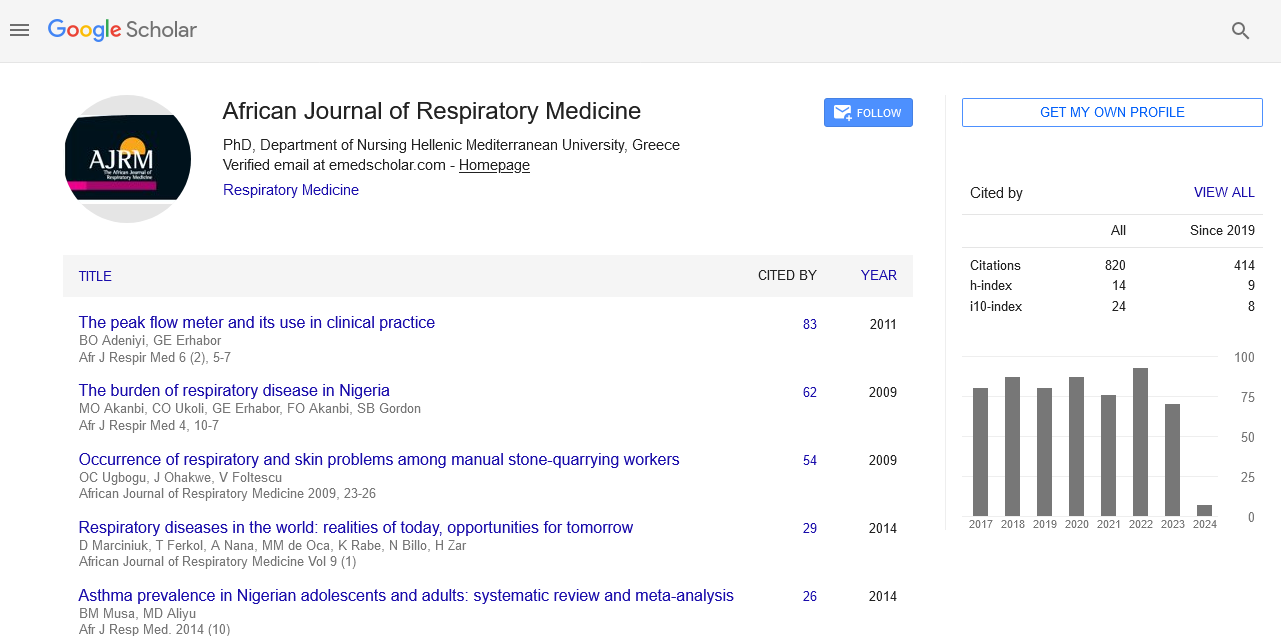
African Journal of Respiratory Medicine received 855 citations as per google scholar report
Research Article - (2025) Volume 20, Issue 1
Received: 02-Oct-2025, Manuscript No. ajrm-25-171569; Editor assigned: 06-Oct-2025, Pre QC No. ajrm-25-171569 (PQ); Reviewed: 21-Oct-2025, QC No. ajrm-25-171569; Revised: 13-Nov-2025, Manuscript No. ajrm-25-171569 (R); Published: 21-Nov-2025, DOI: 10.54931/1747-5597.25.20.62
Introduction: Neuromuscular Electric Nerve Stimulation (NMES) is a modality that is gaining interest as part of the management of participants with Chronic Obstructive Pulmonary Disease (COPD). Given the increased knowledge of how the muscle changes structurally as the respiratory condition deteriorates, treatment options are being investigated to help maintain and improve strength and endurance in patients who are limited due to poor exercise tolerance and increased dyspnoea and cannot part-take in rehabilitation.
Method: Using a randomised control trial design, 33 participants who required admission to hospital due to an acute exacerbation of COPD, with a moderate to severe classification based on the MRC score, were recruited and randomly allocated to an experimental (males: 83.33%, females: 16.67%) or control group (males: 60%, females: 40%). Isometric quadriceps strength was assessed using the hand-held dynamometer, and quadriceps endurance was evaluated with the quadriceps endurance test. Additionally, the rectus femoris cross-sectional area, width, and thickness were measured using an ultrasound. All outcome measures were measured at baseline and upon discharge from the hospital, following the intervention, in all participants. The experimental group received 30 minutes of NMES stimulation on the quadriceps muscles daily throughout their hospitalisation stay (mean days: Experimental – 7.78 days, Control 9.45 days), over and above the usual physiotherapy treatment provided to all participants by physiotherapists in the ward. The control group received only Physiotherapy treatment provided in the ward. All outcome measures were repeated before discharge.
Results: Statistically significant improvements in rectus femoris thickness (left and right: P<0.001) and rectus femoris width on the right side (P=0.017) were reported in the experimental group. In contrast, statistically significant declines in mean values for the left and right Rectus Femoris width were registered for the control group (left: P=0.006, right: P< 0.001).
Conclusion: 30 minutes of daily NMES during a period of hospitalisation due to an AECOPD in moderate to severe participants resulted in significant improvements in all outcome measures for the experimental group, more specifically the quadriceps muscle strength, quadriceps endurance and rectus femoris CSA, width and thickness. Therefore, NMES can be utilised as an adjunct to the already provided physiotherapy treatments offered to maintain and improve quadriceps strength, endurance and muscle structure.
Acute exacerbation COPD; COPD; Chronic obstructive pulmonary disease; NMES; Quadriceps morphology; Ultrasound scan
Chronic Obstructive Pulmonary Disease (COPD) is a progressive respiratory disorder marked by persistent airflow limitation due to lung tissue damage and small airway disease [1]. Beyond lung impairment, COPD causes systemic effects, notably skeletal muscle dysfunction, leading to exercise intolerance and reduced quality of life. COPD patients show a shift in muscle fiber types, with fewer type I oxidative fibers and more type II glycolytic fibers, causing reduced endurance, increased fatigue, and impaired oxidative metabolism [1,2].
Muscle atrophy, especially in the lower limbs, is a key feature of COPD-related muscle dysfunction, worsened by physical inactivity [3-5]. Systemic inflammation and prolonged corticosteroid use further contribute to muscle wasting, reducing strength and increasing energy demands for minimal activity [6,7]. Molecularly, the decline in type I fibers impairs oxidative metabolism, while increased type II fibers reduces endurance and ATP production [2]. Inactivity, malnutrition, and exacerbations accelerate muscle loss, with patients sometimes walking less than ten minutes daily during exacerbations, leading to rapid declines in quadriceps strength [8-10].
To address muscle dysfunction, Neuromuscular Electrical Stimulation (NMES) offers a promising low-metabolic-demand therapy. NMES induces controlled muscle contractions via skin electrodes, benefiting both stable and unstable COPD patients, especially those bedridden during exacerbations [11,12]. It improves muscle structure, exercise capacity, and quality of life [13]. However, research on NMES during acute exacerbations is limited, with most studies focusing on home-based settings and small cohorts [14,15]. No studies contraindicate NMES in acute exacerbations.
As COPD patients struggle with dyspnoea and muscle dysfunction, alternative training modalities that minimise respiratory distress are needed. NMES provides a viable solution by targeting large, fast-twitch motor units, potentially preventing muscle atrophy and improving overall mobility in COPD patients. Recent advancements have improved patient comfort, allowing for stronger yet painless muscle contractions that may be more effective than traditional exercise [16]. Given these potential benefits, the present study aims to investigate changes in quadriceps muscle strength, endurance, and morphological adaptations in moderate to severe COPD patients during hospitalisation for an acute exacerbation.
Study Design and Participants
This randomised single-blinded clinical trial assessed 136 potential candidates referred by respiratory consultants at Mater Dei Hospital, Malta. Of these, 78 met the inclusion and exclusion criteria. A total of 34 participants were assigned to the control group, while 44 were enrolled in the experimental group. All participants provided informed consent. The intermediary physiotherapist identified potential candidates, ensuring blinding of participant allocation. Ethical approval was obtained from the University Research Ethics Committee (UREC) of Malta and the Faculty Research Ethics Committee (FREC) (Code number: FHS-2021-00004). Additionally, the study was registered under ClinicalTrial.gov Identifier NCT05539547.
Randomisation and Masking
Participants were randomly assigned to either the control or experimental group using a pre-generated computer-based randomisation sequence. Allocation was concealed in sequentially numbered, sealed envelopes stored in a locked cupboard. Only the researcher had access to the pre-generated computer-based randomisation sequence.
Patients
Eligible participants were hospital-admitted patients diagnosed with an acute exacerbation of COPD between December 2022 and January 2024. Inclusion criteria consisted of:
• Males or females over 40 years’ old.
•Confirmed diagnosis of acute exacerbation of COPD.
•Medically stable condition.
•Forced Expiratory Volume (FEV) ≤ 50%.
•Medical Research Council (MRC) dyspnoea scoreof 3 or more (moderate to severe).
Exclusion criteria included:
• Mobility or neurological conditions.
• Malignancy.
• Presence of a pacemaker or ImplantableCardioverter Defibrillator (ICD).
• Acute venous thromboembolism.
• Disability or recent orthopaedic surgeries.
Outcomes
Baseline assessments included demographic data, reason for admission, and medical history. Primary outcome measures were isometric quadriceps strength, using a Lafayette Hand-Held Dynamometer (HHD). The patients were to sit on a chair with armrests, the HHD was placed 10 cm above the lateral malleolus, and the patient was asked to extend the knee for a 5-second hold. Three readings were calculated, and an average was taken.
The quadriceps endurance test was used to assess any change in quadriceps endurance throughout hospitalization [17]. The patients were asked to sit on a chair, with both knees and hips at 90°. Both arms crossed over the chest. The patients were asked to extend the lower limb against a weight corresponding to 70% of 1RM at a pace of 12 movements per minute until the point of exhaustion. Secondary outcomes included muscle morphology (width, length, and cross-sectional area) evaluated diagnostic ultrasound at admission and discharge.
Ultrasonographic assessment [18].
Interventions
Control Group (CG): Received standard physiotherapeutic intervention, including chest physiotherapy and mobility exercises.
Experimental Group (EG): Received standard physiotherapeutic intervention with the addition of NMES during hospitalisation for a maximum of 30 days, which was administered by the researcher-physiotherapist.
The parameters used were a frequency of 50 Hz or more, a pulse duration of 400, and a duty cycle of 2 seconds on and 18 seconds off. A duration of 30 minutes per session was carried out, 7 days a week, from admission to discharge, for a period not exceeding 30 days. The intensity used varied for each enrolled patient. This was based on the maximal tolerance of intensity or a visible quadriceps muscle contraction, with the possibility of eliciting slight discomfort. These parameters are based on previous studies since there is no clear guidance on the optimal set of parameters for NMES use in AECOPD
patients.15 A total of 4 electrodes were placed over each of the quadriceps group of muscles, two distal to the hip joint and two proximal to the knee joint (Figure 1).

Figure 1: Electrode positions
Statistical Analysis
Data analysis was performed using SPSS software. Paired t-tests or Wilcoxon signed-rank tests were used for within-group comparisons. In contrast, independent t-tests or Dataanalysis was performed using SPSS software. Paired t-testsor Wilcoxon signed-rank tests were used for within-groupcomparisons. In contrast, independent t-tests or Mann-Whitney U tests were applied for between-groupcomparisons, depending on the normality of the data. A p-value of less than 0.05 was regarded as statisticallysignificant. Quadriceps strength (left and right), quadricepsendurance (right), rectus femoris cross-sectional area (leftand right), and rectus femoris thickness (left and right)were all normally distributed. On the other hand, the leftquadriceps endurance and the rectus femoris width (leftand right) were not normally distributed.
Out of the 78 recruited participants, 15 completed the study duration with the pre-and post-assessment for the control group, while 18 completed the study duration for the experimental group (Figure 2). Forty-five of the participants were excluded: 17 participants lacked compliance or withdrew their consent, and 4 participants had a wrong diagnosis, that is, being admitted with a CHF exacerbation and not a COPD exacerbation, 9 participants developed the infective disease (COVID-19) requiring isolation, 2 participants required CPR, one patient developed lower limb ischaemia and 12 participants were referred late, or discharged soon after referral and initial assessment (Tables 1 and 2).

Figure 2: Enrolment flow chart
| Variables | Control group | Standard deviation | Experimental group | Standard deviation | p-value |
| Age (Years) | 73 (64 -86) | 7.778 | 70 (63-83) | 9.445 | 0.821 |
| Males | 9 (60%) | 15 (83.33%) | |||
| Severity: MRC 3 | 7 (46.67%) | 4 (22.22%) | |||
| MRC 4 | 6 (40%) | 8 (53.33%) | |||
| MRC 5 | 2 (13.33%) | 6 (33.33%) | |||
| Quadriceps strength (kg) Left |
11.08 | 3.193 | 11.65 | 3.867 | 0.65 |
| Quadriceps strength (kg) Right |
11 | 2.569 | 11.45 | 3.646 | 0.692 |
| Quadriceps endurance (Tlim/Seconds) Left | 5.47 | 2.615 | 4.61 | 2.748 | 0.323 |
| Quadriceps Endurance (Tlim/Seconds) Right | 5.47 | 2.279 | 5.11 | 2.541 | 0.702 |
| Rectus FemorisCSA (cm2) Left |
2 | 0.687 | 2 | 1.109 | 0.846 |
| Rectus FemorisCSA (cm2) Right |
3 | 0.923 | 3 | 1.296 | 0.812 |
| Rectus FemorisThickness (mm) Left |
38 | 2.145 | 37 | 2.761 | 0.597 |
| Rectus FemorisThickness (mm) Right |
38 | 2.583 | 38 | 3.189 | 0.976 |
| Rectus FemorisWidth (mm) Left |
7 | 6.601 | 7 | 6.622 | 0.863 |
| Rectus FemorisWidth (mm) Right |
7 | 11.113 | 6 | 10.819 | 0.668 |
| Length of Stay (days) | 9.87 | 7.88 |
Table 1: Mean pre-test values for the control and the experimental groups
|
Control group (n=15) |
Experimental group (n=18) |
|
||||||||
|
|
Admission |
Standard deviation |
Discharge |
Standard deviation |
Admission |
Standard deviation |
Discharge |
Standard deviation |
p-value Control group |
p-value Experimental group |
|
Quadriceps strength (kg) Left |
11.08 |
3.193 |
10.94 |
3.119 |
11.65 |
2.569 |
13.36 |
3.681 |
P=0.252 |
P<0.001 |
|
Quadriceps strength (kg) Right |
11 |
3.867 |
10.99 |
2.584 |
11.45 |
3.646 |
13.43 |
3.074 |
P=0.974 |
P<0.001 |
|
Quadriceps endurance (Tlim/Seconds) Left |
4.75 |
2.615 |
3 |
3 |
2.5 |
2.279 |
4.79 |
4.79 |
P=0.380 |
P=0.027 |
|
Quadriceps endurance |
5.47 |
2.748 |
5.67 |
2.664 |
5.11 |
2.541 |
6.06 |
2.754 |
P=0.486 |
P=0.015 |
|
Rectus femorisCSA (cm2) Left |
2.41 |
0.687 |
2.15 |
0.717 |
2.48 |
1.109 |
2.91 |
1.18 |
P=0.186 |
P=0.012 |
|
Rectus femorisCSA (cm2) Right |
2.71 |
0.923 |
2.62 |
0.694 |
2.81 |
1.296 |
3.38 |
1.392 |
P=0.603 |
P=0.003 |
|
Rectus FemorisThickness (mm) Left |
6.85 |
2.145 |
6.27 |
2.181 |
6.71 |
2.583 |
7.78 |
2.955 |
P=0.118 |
|
|
Rectus FemorisThickness (mm) Right |
6.91 |
2.761 |
7.08 |
2.414 |
6.46 |
3.189 |
7.93 |
3.781 |
P=0.641 |
P<0.001 |
|
Rectus FemorisWidth (mm) Left |
38.3 |
6.601 |
34.73 |
-2.731* |
26.56 |
11.113 |
37.59 |
-1.846* |
P= 0.006 |
P=0.065 |
|
Rectus FemorisWidth (mm) Right |
37.8 |
6.672 |
35.96 |
-3.408* |
37.9 |
10.819 |
40 |
-2.391* |
P<0.001 |
P=0.017 |
Table 2: Mean comparison of pre-test vs. post-test for quadriceps strength, endurance and ultrasound scans of the rectus femoris muscle
Isometric Quadriceps Strength
Neuromuscular Electrical Stimulation (NMES) significantly improved isometric quadriceps strength in participants with COPD. The experimental group demonstrated statistically significant increases of 14.68% in left quadriceps strength (p<0.001), with mean values rising from 11.65 kg pre-intervention to 13.36 kg post-intervention (Figure 2). In contrast, the control group exhibited a minor, non-significant decline of 1.26% (p=0.252), from 11.08 kg to 10.94 kg (Figure 3).
Similarly, for the right quadriceps, the experimental group saw a 17% improvement (p<0.001), with strength increasing from 11.45 kg to 13.43 kg post-intervention. The control group, however, showed negligible change (-0.01 kg, p=0.974) (Figure 3). These findings highlight NMES’s ability to activate more motor units and facilitate neural plasticity, contributing to strength gains even in immobilised COPD patients.
Quadriceps Endurance
NMES intervention also significantly enhanced quadriceps endurance. The experimental group showed a 0.72TlimQ-second increase in left quadriceps endurance (p=0.027), improving from a baseline of 4.61TlimQ seconds to 5.33TlimQ seconds post-intervention (Figure 3). In contrast, the control group experienced a decline of -0.27TlimQ seconds, but this change was not statisticallysignificant (z=-0.877, p=0.380).

Figure 3: Mean change in quadriceps strength (kg) and endurance (Tlim/s) between the control and experimental groups for the left and right groups of muscles.
For the right quadriceps, NMES led to a statistically significant improvement of 0.95TlimQ seconds (p=0.015), increasing endurance from 5.11TlimQ to 6.06TlimQ seconds (Figure 4). The control group exhibited only a minor increase of 0.2TlimQ seconds (3.66% improvement), which was not statistically significant. The experimental group demonstrated a markedly higher percentage improvement of 18.60%, reinforcing NMES’s effectiveness in enhancing muscle endurance in COPD patients.
Rectus Femoris Cross-Sectional Area (RF CSA) and Thickness
A statistically significant increase in rectus femoris cross-sectional area (RF CSA) was observed in the experimental group, with a 17% improvement in the left RF CSA (p=0.012) and a 20% increase in the right RF CSA (p=0.003 (Figure 4). Mean values rose from 2.48 cm2 to 2.91 cm2 (left) and from 2.81 cm2 to 3.38 cm2 (right). Conversely, the control group experienced an 11% reduction in left RF CSA (from 2.41 cm2 to 2.15 cm2) and a 3% reduction in right RF CSA (from 2.71 cm2 to 2.62 cm2).
Rectus femoris thickness also improved significantly in the experimental group, with a 16% increase on the left and a 23% increase on the right (p<0.001) (Figure 4). The control group, however, exhibited a non-significant decline of 8.5% on the left (p=0.118) and a marginal, non-significant 3% increase on the right.
Rectus Femoris Width (RF Width)
The control group showed a statistically significant deterioration in rectus femoris width (p=0.006 on the left, p<0.001 on the right). The left RF width in the control group declined by 9%, from 38.3 mm to 34.73 mm, whereas the right RF width dropped from 37.8 mm to 35.96 mm (Figure 4). In contrast, the experimental group exhibited a 3% increase in left RF width, though it did not reach statistical significance (p=0.186). However, the right RF width significantly increased from 37.90 mm to 40 mm (p=0.017), reinforcing the positive morphological effects of NMES.

Figure 4: Mean change in rectus femoris cross-sectional area (cm2), width and thickness (mm) between the control and experimental groups for the left and right groups of muscles
The findings of this study underscore the potential of NMES as a therapeutic intervention for patients with moderate to severe Chronic Obstructive Pulmonary Disease (COPD) during acute exacerbations. By targeting quadriceps muscle strength, endurance, and morphology, NMES offers a promising alternative to conventional exercise regimens, particularly for individuals with limited mobility due to severe dyspnoea. The observed statistically significant improvements in muscle strength and endurance highlight NMES's efficacy in mitigating muscle deterioration, which is critical given the association between quadriceps weakness and poor prognosis in COPD patients [19].
Previous research has demonstrated that NMES can enhance muscle strength and exercise capacity in COPD patients, particularly in those who are unable to engage in conventional exercise training due to severe respiratory symptoms. A systematic review and meta-analysis by Alves et al. concluded that NMES improves exercise capacity and muscle strength in individuals with COPD,suggesting that it should be incorporated into pulmonary rehabilitation programs [20]. This aligns with findings from Vivodtzev et al., who reported that NMES led to significant quadriceps hypertrophy and improved exercise tolerance, even in patients with advanced COPD [21].
Similarly, a Randomised Controlled Trial (RCT) by Maddocks et al. found that NMES improved functional exercise capacity in patients with severe COPD by enhancing quadriceps muscle mass and function [22]. Their findings indicate that NMES not only prevents muscle atrophy but also facilitates muscle regeneration, possibly through enhanced neural activation and increased muscle fiber recruitment. These physiological adaptations are particularly relevant in COPD patients, as muscle atrophy is a well-documented consequence of systemic inflammation, corticosteroid use, and prolonged inactivity [4,6].
However, the present study's focus on hospitalised patients during acute exacerbations is noteworthy. While NMES has been widely studied in stable COPD patients, its application during acute exacerbations has been less explored. This study contributes new insights by demonstrating that even short-duration NMES intervention during hospitalisation can lead to measurable improvements in muscle strength and endurance. These findings suggest that early NMES implementation may help counteract the rapid muscle deterioration seen during exacerbations, potentially reducing hospital stay duration and improving post-discharge recovery [23].
The observed improvements in muscle strength and endurance align with findings from other studies. For instance, Dal Corso et al. reported that NMES led to increased type II muscle fiber cross-sectional area in moderately impaired COPD patients, indicating potential for muscle hypertrophy and strength gain. These adaptations may result from NMES's ability to stimulate high-threshold motor units, which are often underutilised in COPD patients due to disuse atrophy and metabolic inefficiency [24].
Additionally, the structural muscle adaptations observed in this study such as increased Rectus Femoris Cross-Sectional Area (RF CSA) and muscle thickness—are consistent with findings from McKay et al., who reported muscle hypertrophy and improved oxidative metabolism following NMES in patients with muscle-wasting conditions. These morphological improvements are particularly significant given that muscle atrophy in COPD is associated with decreased survival and functional impairment [25].
Furthermore, while traditional exercise-based rehabilitation remains the gold standard for improving muscle function in COPD, many patients experience exercise intolerance due to severe dyspnoea, fatigue, and cardiovascular limitations Maltais et al. NMES provides a low-metabolic-demand alternative, allowing patients to maintain muscle activity without exacerbating respiratory distress. Studies by Sillen et al. and Vivodtzev et al.
support this notion, highlighting NMES's ability to improve functional performance and increase muscle oxidative capacity, even in sedentary and severely deconditioned COPD patients.
Collectively, these findings suggest that NMES is a viable adjunctive therapy for COPD patients, particularly those unable to engage in traditional exercise due to acute exacerbations or severe mobility impairments. The present study further supports the integration of NMES into comprehensive rehabilitation programs, particularly in hospitalised settings, where early muscle stimulation may prevent exacerbation-related muscle atrophy. Given the growing evidence supporting NMES, future research should focus on optimising stimulation parameters, exploring long-term benefits, and investigating potential synergistic effects when combined with pharmacological and nutritional interventions [26,27].
The results discussed above need to be interpreted with caution, mostly because of the small population sample. The small population sample reduces the statistical power of the results obtained; therefore, there was less power to detect true effects and an increased likelihood of type 2 errors. Similarly, these results could have been susceptible to outliers, which can skew the results with wider confidence intervals. Furthermore, the small population size cannot be a representative figure of the broader populations, enhancing the risk of bias, more specifically, selection bias.
The results demonstrate that NMES significantly enhances quadriceps strength, endurance, and muscle morphology in COPD patients hospitalised due to acute exacerbations. While the control group exhibited muscle deterioration or negligible changes, the NMES intervention led to substantial improvements, particularly in quadriceps muscle strength, quadriceps enduranc and rectus femorisCSA and thickness. These findings suggest that NMES is an effective adjunct to conventional rehabilitation strategies for COPD patients at risk of muscle atrophy.
This research received no specific grants from any public, commercial, or not-for-profit agencies.
RD thanks Ms. Geraldine Cremona for her assistance with patient identification, referral, and recruitment of participants. Special appreciation is also extended to Dr. Kieran Chircop (Consultant Radiologist) and Ms. Francesca Pace-Bonello for their contributions in scheduling and performing ultrasound scans throughout the study.
The authors declare no conflict of interest.
[Crossref] [Google Scholar] [PubMed]
[Crossref] [Google Scholar] [PubMed]
[Crossref] [Google Scholar] [PubMed]
[Crossref] [Google Scholar] [PubMed]
[Crossref] [Google Scholar] [PubMed]
[Crossref] [Google Scholar] [PubMed]
[Crossref] [Google Scholar] [PubMed]
[Crossref] [Google Scholar] [PubMed]
[Crossref] [Google Scholar] [PubMed]
[Crossref] [Google Scholar] [PubMed]
[Crossref] [Google Scholar] [PubMed]
[Crossref] [Google Scholar] [PubMed]
[Crossref] [Google Scholar] [PubMed]
[Crossref] [Google Scholar] [PubMed]
[Crossref] [Google Scholar] [PubMed]
[Crossref] [Google Scholar] [PubMed]
[Crossref] [Google Scholar] [PubMed]
[Crossref] [Google Scholar] [PubMed]
[Crossref] [Google Scholar] [PubMed]
[Crossref] [Google Scholar] [PubMed]
[Crossref] [Google Scholar] [PubMed]
[Crossref] [Google Scholar] [PubMed]
[Crossref] [Google Scholar] [PubMed]
[Crossref] [Google Scholar] [PubMed]
[Crossref] [Google Scholar] [PubMed]
[Crossref] [Google Scholar] [PubMed]
Select your language of interest to view the total content in your interested language
To read the issue click on a cover