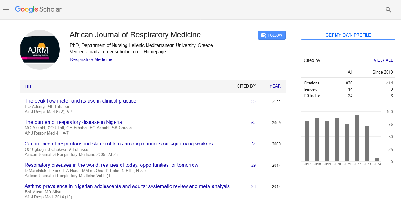
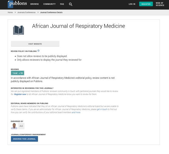
African Journal of Respiratory Medicine received 855 citations as per google scholar report
Mini Review - (2023) Volume 18, Issue 2
Received: 01-Mar-2023, Manuscript No. ajrm-23-90569; Editor assigned: 03-Mar-2023, Pre QC No. ajrm-23-90569 (PQ); Reviewed: 17-Mar-2023, QC No. ajrm-23-90569; Revised: 22-Mar-2023, Manuscript No. ajrm-23-90569 (R); Published: 29-Mar-2023, DOI: 10.54931/1747 5597.22.18.80
Background: Physiotherapy plays a major role in the long-term management of COPD. Guidelines issued by the BTS (2013) and the GOLD guidelines (2017) set a strong recommendation for pulmonary rehabilitation, chest mobility and breathing exercises as a management for these patients. Research is now focusing on METs as an adjunct to other treatments but evidence is still inconclusive. The aim of this review is to evaluate the effectiveness of METs on chest expansion, pulmonary function and exercise tolerance for patients diagnosed with COPD.
Design: Narrative review of research studies.
Data sources: Google Scholar, Pubmed, Medline, Embase and Research Gate were searched for all journal articles published between 2000-2023.
Method: Papers were included under the population intervention comparator outcome (PICO) framework. Critical appraisal, data abstraction, and synthesis were carried out by the different authors.
Results: Out of 23 titles screened, 7 interventions with a low to moderate risk of bias met the inclusion criteria. Studies that implemented METs intervention were observed to have promising results on chest expansion, pulmonary function and exercise tolerance when integrated with self-stretching exercises, chest mobility exercises and/or breathing exercises, aerobic training and pulmonary rehabilitation.
Conclusion: METs as an adjuvant treatment in patients with COPD show favourable improvements in pulmonary function, chest expansion and exercise tolerance. However, there is still insufficient evidence.
Impact: Possible positive effects following METs intervention as an adjunct to other physiotherapy treatments may lead to greater health benefits.
Chronic obstructive pulmonary disease; Physiotherapy, Pulmonary hyperinflation
COPD is a progressive and irreversible respiratory condition characterised by expiratory flow limitation which results in air trapping and pulmonary hyperinflation. This leads to musculoskeletal alterations, forcing the diaphragm and intercostal muscles to operate in a mechanically disadvantageous position. For the diaphragm to operate in an optimal manner, its muscle fibers must be able to progressively overlap so as to form cross bridges which increase their contraction force. Following contraction of the diaphragm, cross bridges are broken so that the muscle can return to its dome-shaped position. As a result of the diaphragm being in a shorter and more flattened position, an adequate contractile force is diminished. Following these chronic adaptations, respiratory muscles work at their full capacity to meet the ventilator demands, increasing the work of breathing, resulting in recruitment of the accessory muscles of respiration in compensation. The accessory muscles of respiration adapt to this increased workload by shortening and becoming overactive, resulting in limited upper limb and cervical mobility. This may result in the patient experiencing shortness of breath. Moreover, changes in chest wall rigidity and muscular function may cause postural changes and alterations in rib cage configuration which can result in increased thoracic kyphosis.1-4
Muscle Energy Techniques (METs) are being considered as adjunctive tools in the management of patients with COPD, but their effectiveness has not been well evaluated.
Background
METs are a type of manual therapy used to improve joint mobility, stretch tight muscles and fascia, improve circulation, and decrease pain. During the application of METs, an isometric contraction targeting the tight muscle is performed, activating the inhibitory Golgi Tendon Organs within the same muscle.5,6
Studies are investigating the use of METs as an adjunct to other treatments such as self-stretching exercises, chest mobility exercises and/or breathing exercises, aerobic training and pulmonary rehabilitation to help manage the over-activation of accessory muscles of respiration. By increasing muscle length and range of motion of these accessory muscles, a decrease in thoracic wall stiffness was observed by Clarke et al. leading to improvements in Forced Expiratory Volume in 1 second (FEV1), reduction in respiratory rate and increase in oxygen saturation. Unlike any other manual therapies that assists in muscle lengthening such as passive stretching, high velocity manipulation and massage, METs requires the active participation of the patient. Moreover, it is a safe method which avoids minor or major adverse effects, with the most serious potential side effect of high velocity manipulation being neurovascular complications.1-3,7,8
Therefore, seeing the information above, this review aims at critically analysing literature which investigates the application of METs with another physiotherapy treatment focusing mainly on changes in pulmonary function, chest expansion and exercise tolerance as primary outcome measures.
Aims
This review aims to determine if METs intervention when used as an adjunct to other physiotherapy interventions result in improvements in pulmonary function, chest measurement and functional endurance. The research question therefore states ‘Are METs effective at improving pulmonary function, exercise tolerance and chest expansion in persons living with COPD?’
Search methods
A narrative review design was chosen in this study allowing for a more comprehensive research.
The review used the population (P), intervention (I), comparator (C) and outcome (O) (PICO) framework to guide inclusion criteria and report the study characteristics. Papers retrieved for this review were obtained following a thorough search using Google Scholar, PubMed, Medline, Research Gate and Embase databases.
Table 1 illustrates the key PICO search terms. Data was extracted and critically appraised using a critical appraisal skills programme (CASP tool) to facilitate quality research appraisal. The following information was extracted throughout this process: characteristics of the study (participants and sample size), intervention and control group treatment, and results.
| Population | Intervention | Comparison | Outcome |
|---|---|---|---|
| Adults with COPD | MET intervention | METs compared to self-stretching | Pulmonary function |
| Adults with decreased pulmonary function and chest expansion as a result of thoracic surgery | METs compared to chest mobility exercises | Chest measurement | |
| METs compared to breathing exercises | Functional endurance | ||
| METs compared to breathing and chest mobility exercises | |||
| METs compared to aerobic training | |||
| METs compared pulmonary rehabilitation |
Search strategy
The search strategy yielded 1,092 research articles following a preliminary search through the selected databases. After removal of duplicates (n=614) and of irrelevant studies (n=455), 23 full-text randomized-control trial with pretest and post-test experimental design articles were kept as potentially relevant after title screening. Studies were then screened as per the inclusion and exclusion criteria (Table 2).
| Criterion | Inclusion | Exclusion |
|---|---|---|
| Language of article | Published in English | Published in another language other than English |
| Publication date | Study published less than 20 years ago | Study published more than 20 years ago |
| Statistical method used | Description of the statistical method used is present | Description of the statistical method used is not present |
| Patient selection | Information on patient selection needed to be given, with patient not having an acute illness or recent exacerbation of COPD | Information on patient selection was not provided or study included patients with acute illness or recent exacerbation of COPD |
| Age | Minimum of 18 years old | Sample included individuals younger than 18 years |
| Study design | Randomised control trial with pre-test and post-test experimental design | Systematic Review, Meta-analysis, Non-randomised control trial, Randomised control trial with another experimental design |
| Outcome Measures | Study examined one or more of the following: Lung function Chest measurements Functional endurance capacity | Does not assess any of the primary outcome measures |
| Intervention period and follow up period | The acute effects of MET was investigated in the study with no limitation on follow up period | The chronic effects of MET was investigated with the intervention process being longer than 4 weeks |
Study selection and quality appraisal
A total of 7 articles were then accepted for this narrative review. Tables 3 and 4 provide a summary of 7 articles discussing the effects of METs as adjuncts to self-stretching exercises, chest mobility exercises, breathing exercises, breathing and chest mobility exercises, aerobic training and pulmonary rehabilitation.
| Author and Year of Publication | Title | Research Design and Data Collection Instrument | Results | Strengths and Limitations |
|---|---|---|---|---|
| Anand, Narwal and Sindhwani (2013). | Accessory Inspiratory Muscles Energy Technique effect on Pulmonary Function in COPD Subjects. | Randomized control trial. Tests carried out: Chest expansion, Dyspnea, Exercise tolerance, Respiratory rate, Heart rate, Oxygen saturation and Quality of life. |
When analysing chest expansion measurements the intervention group registered improvements in chest expansion from 1.56 cm at baseline to 2.59 cm post-intervention (p=0.001), whereas the control group had less of an improvement with 1.39 cm at baseline to 1.95 cm post-intervention. 6MWT values were also noted to increase significantly by 226 m in the intervention group when compared to 91 m registered by the conventional group (p=0.001). | A control group was recruited thereby increasing the reliability of the study. Limited description of the intervention carried out was provided, therefore making it difficult to replicate the study and assess for reliability. |
| Dave, Thaker and Gondaliya (2019). | A Study to find out the effect of Accessory Inspiratory Muscle Energy Technique on Chest Expansion and Pulmonary Function in an Elderly Male. |
Experimental study Tests carried out: Pulmonary function tests and Chest expansion. |
The MET group was noted to have had statistically significant improvements in chest expansion measures with a total of 0.29 cm and 0.32 cm increase at the axillary and xiphesternal levels. The chest mobility exercises group were only noted to have minimal improvements of 0.1 cm and 0.2 cm respectively. Statistical significant difference was observed when comparing the control group with intervention both at axillary (p=0.001) and xiphesternal (p=0.002) level. The improvements in chest expansion for the MET group were also accompanied with changes in pulmonary function measures. A statistically significant improvement in FVC (0.37 L [p=0.003]) and FEV1 (0.41 L [p=0.001]) were reported. Though an improvement in FEV1/FVC ratio (0.92%) was observed it was not statistically significant (p=0.221). Pulmonary function measures in the control group improved by a lesser degree (0.14 L, 0.16 L and 0.04% respectively). | A detailed explanation of both the intervention and the outcome measure performed was provided. A follow-up period was not considered in this study and the kyphotic curvature was not measured. |
| Krishna et al. (2018). | Study to Find out the Efficacy of Osteopathic Manual Therapy in Chest Expansion in COPD Patients. | Randomized control trial. Tests carried out: Chest expansion. |
The mean values at axillary level, 4th intercostal space level and xiphisternal level showed improvements from pre-intervention measurements to those at 30 minutes post-intervention for both groups. When observing each level individually, measures at the axillary level for the MET group resulted in a total percentage increase of 21% (p=0.001) from 1.9 cm at baseline to 2.3 cm 30 minutes after the intervention, whereas the aerobic exercises group also reported an improvement but only of a 10% from 2.08 cm at baseline to 2.29 cm (p=0.034) post-intervention. The MET group then registered after 24 hours following intervention with a slight decline in scores of a total percentage decrease of 4% (p=0.01) at 24 hours post the intervention and the aerobic exercises group also had a decrease in the total percentage score of 33% (p=0.135) 24 hours after. Results taken at the 4th intercostal space show that the MET group registered a superior improvement of 29% (p=0.001) from 1.74 cm expansion pre-intervention to 2.25 cm 30 mins post-intervention, a value which declined to a percentage value of 14% to 2.11 cm (p=0.001) 24 hours after. In comparison, the aerobic exercises group had a total increase of 22% (p=0.001) from 1.70 cm to 2.07 cm a value which decreased to a percentage value of 5% to 2.02 cm (p=0.003) 24 hours post-intervention. Lastly, at the xiphisternal level, the MET group measured an improvement of 39% (p=0.001) from 1.66 cm at baseline to 2.30 cm at 30 mins after the intervention and a further increase of 46% (2.42 cm) (p=0.001) at 24 hours post-intervention. The aerobic exercises group however improved by 38% (1.75 cm pre-intervention and 2.42 cm 30 mins post-intervention [p=0.001]) a value which dropped to 1.82 cm (p=0.586) at 24 hours post-intervention at the same level. | A large sample population was used and a control group was included. Moreover, a detailed explanation of the exercise methodology was provided. Only male participants of a relatively young age that is 25-55 years suffering from chronic lung disease were recruited. Limited information on the demographics of the population such as smoking cessation, severity of COPD, age percentage distribution of the patients recruited was not provided. |
| Rakesh and Jibi (2016). | The Effectiveness Of Post Isometric Relaxation Technique In Relation With Pulmonary Function And The Chest Expansion Of Post Thoracic Surgery Patients. | Randomized control trial. Tests carried out: Chest expansion measurement, Forced vital capacity Forced expiratory volume in 1 second. |
Statistically significant improvements in FVC were observed in both groups but the MET group registered a higher percentage improvement from baseline values of 48.39% (p<0.001), as compared to the breathing exercise group who had an improvement of 40.85% (p<0.016). This was also noted in the FEV1 measures with the MET group having a total percentage improvement of 27.89% from baseline (p<0.001) as compared to a total percentage improvement of 19.57% (p<0.016) for the latter group. Moreover, statistically significant improvement (p<0.001) at axillary, nipple and xiphoid levels were observed on comparison of pre-test and post-test in the intervention group (pre-test; axillary level-M-1.6 ± 0.13 cm; nipple level-M-3.26 ± 0.14 cm; xiphoid level-5.88 ± 0.37, post-test-axillary level-M-2.33 ± 0.38 cm; nipple level-M-4.55 ± 0.2 cm; xiphoid level-6.93 ± 0.35 cm). On the other hand statistically significant decrease was observed at nipple (p<0.026) and xiphoid levels (p<0.001) on comparison of pre-test and post-test in the control group (pre-test; nipple level-M: 3.38 ± 0.15 cm; xiphoid level-M-5.27 ± 0.14 cm, post-test nipple level-M: 3.23 ± 0.13 cm; xiphoid level-M-6.11 ± 0.44 cm). Axillary level in the control group further decreased following post-test measurements (pre-test: M-1.5 ± 0.15 cm, post-test; M-1.3 ± 0.15 cm) however did not reach statistical significance (p>0.06). |
A detailed description of the MET intervention on pectoralis major and minor was given, thereby increasing the validity of the study. A control group was included enabling a better comparison of the effects brought about by the post-isometric intervention. Long term effects of the intervention were not investigated. The assessor was not blinded to the participants therefore measurement bias would need to be considered. |
| Sule, Kakade and Palekar (2017). | Effect of Static Stretching And Pulsed MET on Accessory Muscles in COPD Patients: A Comparative Study. | Randomized control trial. Test carried out: Forced expiratory volume in 1 second, Respiratory rate and Oxygen saturation. |
Pulsed METs group showed statistically significant improvements (p=0.001) in all the outcome measures with the MET group resulting in greater improvement of 6.13% from baseline (baseline-FEV1 % predicted: 43.27%, post-intervention-FEV1 % predicted: 49.4%). Participants in the static stretching group on the other hand reported a non-significant improvement of 0.06% (44.47% FEV1 % predicted at baseline to 44.53% FEV1 % predicted post-intervention). |
The sampling age group was between 40-60 years old; with both males and females recruited therefore it can be broadly reflected to the COPD population. The authors did not provide a detailed description of the intervention used and in which position was pulsed MET performed. |
| Wada et al. (2016). | Effects Of Aerobic Training Combined With Respiratory Muscle Stretching On The Functional Exercise Capacity And Thoracoabdominal Kinematics In Patients With COPD: A Randomized And Controlled Trial. |
Randomized control trial. Tests carried out: 6 Minute Walk test, Optoelectronic plethysmography and Surface electromyography. |
The treatment group had a mean average value of 473 m during a 6-minute walk test at baseline. The control group had a mean average value 439 m during a 6-mintue walk test at baseline. Post intervention, groups improved equally in their walking distance by 15 m (p<0.001). Therefore, the authors did not report a difference in their exercise tolerance between the experimental group and the control group. | The duration of the study was that of 12 weeks, with 2 sessions conducted per week. Furthermore, a control group was recruited for better comparison of the intervention. The stretching protocol integrated 2 techniques; METs and passive stretching. Therefore, it is not possible to determine which technique was more effective in causing the resultant benefits. Moreover, initial maximal exercise testing was not performed; rather 25% of maximal exercise intensity was performed, which might not be equally suitable for all patients. |
| Zanotti et al. (2012). | Osteopathic Manipulative Treatment Effectiveness In Severe Chronic Obstructive Pulmonary Disease: A Pilot Study. |
Longitudinal study. Tests carried out: Pulmonary function and Exercise capacity. |
The authors concluded that the addition of manual therapy intervention to Pulmonary Rehabilitation resulted in an increase in chest wall mobility which led to a decrease in residual volume with a difference of 0.5 L (p=0.05) from pre to post-intervention. This led to an improvement in exercise capacity with a difference of 48.8 m observed between the 2 groups. The control group increased their exercise tolerance by 23.7 m post-intervention and the experimental group had a total increase of 72.5 m post-intervention. A difference of 48.8 m was therefore observed between the 2 groups following intervention which was statistically significant (p=0.04). | Detailed information on the demographic of the sample population was provided. Moreover, the intervention consisted of 5 days a week for 4 weeks therefore allowing effects of the intervention to take place. The clinicians that performed the osteopathic intervention were not experienced osteopathic practitioners but rather students from the School of Osteopathic Manipulation. Therefore lack of experience in the delivery of the treatment may have caused variation in treatments. Moreover, the small size limits generalization of the results. |
| Author and Year of Publication | Population information and sample size | Intervention Group | Control Group | Confounding factors |
|---|---|---|---|---|
| Anand, Narwal and Sindhwani (2013). | Patients suffering from moderate to severe COPD. Mean age for intervention group-61.20 ± 5.84 years Mean age for control group-61.53 ± 7.94 years Sample size-30 (Females-3, Males-27) |
MET intervention and conventional chest physiotherapy (diaphragmatic breathing, pursed lip breathing, postural drainage and thoracic expansion exercises). | Conventional chest physiotherapy (diaphragmatic breathing, pursed lip breathing, postural drainage and thoracic expansion exercises). | |
| Dave, Thaker and Gondaliya (2019). | Elderly male with no previous diagnosis of respiratory conditions. Distribution of different age group: 65-68 years-19 participants 69-71 years-9 participants 72-75 years-2 participants Sample size-30 males |
MET intervention with chest mobility exercises. | Chest mobility exercises. | Kyphotic curvature of participants was not measured which may have influenced their lung function parameters. |
| Krishna et al. (2018). | Males between the ages of 25-55 years suffering from a minimum of 1 year history of chronic lung disease. Sample size-105 males |
Osteopathic manual therapy (including MET intervention) with conventional physiotherapy (abdominal exercises, incentive spirometry, upper and lower extremity exercises and aerobic exercises) and components of pulmonary rehabilitation (smoking cessation, clearing of secretion, relaxation positions, oxygen therapy, energy conservation techniques, fatigue obviation mechanisms, pranayama, nutritionary guide lines) | Conventional physiotherapy (abdominal exercises, incentive spirometry, upper and lower extremity exercises and aerobic exercises and components of pulmonary rehabilitation (smoking cessation, clearing of secretion, relaxation positions, oxygen therapy, energy conservation techniques, fatigue obviation mechanisms, pranayama, nutritionary guide lines) | Components of rehabilitation may have the improvements in both groups. |
| Rakesh and Jibi (2016). | Participants who underwent postero-lateral thoracotomy and were stable on the 4th day of surgery. Mean age of intervention group-55 ± 4.17 years Mean age of control group-53.16 ± 4.17 years Sample size-30 (Females-9, Males-21) |
MET intervention with diaphragmatic breathing exercise and pursed lip breathing exercises with coughing and lower range of motion exercise. | Diaphragmatic breathing exercise and pursed lip breathing exercises with coughing and lower range of motion exercise. | |
| Sule, Kakade and Palekar (2017). | Patients withs stable grade II and III COPD. Mean age of intervention group-20% for 40-50 years and 80% for 51-60 years Mean age of control group-53.33% for 40-50 years and 46.66% for 51-60 years. Sample size-30 (Females-?, Males-?) |
Pulsed MET and conventional exercises (thoracic expansion exercises, segmental breathing exercise, diaphragmatic breathing exercises). | Static stretching and (thoracic expansion exercises, segmental breathing exercise, diaphragmatic breathing exercises). | |
| Wada et al. (2016). | Patients diagnosed with moderate to severe COPD. Mean age of intervention group-61 ± 5.4 years Mean age of control group-64 ± 5.6 years Sample size-30 (Females-?, Males-?) |
MET intervention and passive stretching techniques with aerobic exercises. | Upper and lower limb muscle stretching with aerobic exercises. | |
| Zanotti et al. (2012). | Stable stage III COPD participants, who did not show signs of exacerbation from at least 3 months. Mean age of intervention group-64.2 ± 5.5 years Mean age of control group-63.5 ± 4.7 years Sample size-20 (Females-5, Males-15) |
Pulmonary rehabilitation with osteopathy treatment (including MET intervention). | Pulmonary rehabilitation with sham osteopathy treatment. | Lack of familiarity to the treatment by the student practitioner may influence results of outcome measures. |
Outcome measures used in these interventions
The identified studies focused on 3 outcome measures being pulmonary function, chest measurements and functional endurance capacity. FEV1 was investigated in 4 studies [Dave, Thaker and Gondaliya, 2019 (P=0.001), Sule, Kakade and Palekar, 2017 (P=0.001), Rakesh and Jibi, 2016 (P<0.001), Zanotti et al., 2012 (P>0.5)], Forced vital capacity (FVC) was investigated by 3 studies [Dave, Thaker and Gondaliya, 2019 (P=0.003), Rakesh and Jibi, 2016 (P<0.001), Zanotti et al., 2012 (P>0.5)], FEV1/FVC ratio was measured in only 1 study (Dave, Thaker and Gondaliya, 2019 (P=0.221)) and another study Zanotti et al., 2012 used residual volume (P=0.05) as outcome measures. Three of the 4 studies observed statistically significant improvements when combining METs intervention to other physiotherapy treatments in FEV1 and FVC, with FEV1/FVC ratio and residual volume. Vital capacity was also reported to have more marked statistical improvements for the intervention group when compared to the control group; however results were not statistically significant, with the author not providing p values.3,9-11
Four studies [Dave, Thaker and Gondaliya, 2019 (P=0.001), Krishna et al., 2018 (P=0.001), Rakesh and Jibi, 2016 (P<0.001) and Anand, Narwal and Sindhwani, 2013 (P=0.001)] looked at changes in chest measurements prior and following METs intervention. Both the control group who received conventional physiotherapy treatment and the intervention group, who received METs intervention together with conventional treatment, resulted in improvements in outcome measurements but the intervention group had statistically significant improvements.9,10,12
Functional endurance capacity was then investigated in 3 papers [Wada et al., 2016 (P<0.001), Anand, Narwal and Sindhwani, 2013 (P=0.001), Zanotti et al., 2012 (P=0.04)]. All 3 studies performed the 6-minute walk test (6MWT) as an outcome measure. Greater statistically significant improvements were observed for the intervention group in all the 3 studies when compared to the control group.11-13
Critical evaluation of articles
All studies included had an unclear risk of bias and limitations in reporting quality when assessed using the Cochrane- risk-of-bias 2.0 tool (Cochrane RoB 2) for randomised trials. Four studies reported adequate random sequence generation; another study did not provide detail on patient selection, while the other 2 studies used purposive sampling method. Two studies used an appropriate method of allocation concealment, while the other studies were judged at unclear risk of bias because they omitted details relating to allocation concealment. In six of the included studies, blinding of participants and personnel was not described despite the use of a sham control, making it unclear whether the lack of blinding would influence outcomes. Only one study specified that assessors of outcome measures were blinded. All of the studies had published protocols, therefore, the selective outcome reporting domain was judged at a low risk of bias (Figures 1 and 2).

Figure 1. Cochrane Risk-of-Bias Tool

Figure 2. Summary of Risk of Bias
This review provides information on the effects of METs in combination to physiotherapy, results which show favourable improvements in pulmonary function, chest measurements and exercise capacity in patients suffering from COPD. Previous systemic reviews focused on investigating the effect of manual therapy including massage, high velocity manipulation and rib cage mobilisation on pulmonary function and exercise capacity or the effects of METs on lung function and exercise capacity as outcome measures. As a result of the small number of studies, limited sample size, difference in METs treatment and methodology, and unclear risk of bias, potential improvements reported should be considered with caution.7,14
Improvements in lung function measures following MET interventions have been attributed to lengthening of the accessory muscles. Modifications to the muscle tissue properties were observed through an increase in sarcomere size and muscle viscoelasticity. Therefore, by reducing muscular, ligament and fascial tonicity to the cervical and thoracic musculature, improvements in the passive component of expiration in patients diagnosed with COPD was reported. This is because of a reduction in the inspiratory block sustained following alteration in ribs position. Further information about patient demographics, methods of analysis and comparison of results would have improved the validity and reliability of outcomes from studies by Sule, Kakade and Palekar (2017) and Rakesh and Jibi (2017). Knowing the patient’s age, severity of their condition and smoking history allows for the identification of which patients would benefit most from such techniques and whether there are any influential factors which might possibly impact outcomes. Moreover, such information would help determine if the participants in a study are a representative sample of a particular population leading to generalisation.1,3,10,15
METs are also reported to lead to an increase in the muscle viscoelasticity and accommodation of the muscle spindle, factors which reduce muscle stiffness, improve muscle performance, and increase thoraco-abdominal mobility. Wada et al. (2016) further reinforce that stretching helps modify thoraco-abdominal mobility better when compared to aerobic training. The METs group showed an increase in lung volume, which is associated with a greater abdominal contribution.3,14
Apart from improvements in lung function measures, the integration of METs and chest mobility exercises resulted in significant improvements in chest expansion. Dave, Thaker and Gondaliya (2019) reported that these results were due to the attainment of an adequate length in the accessory muscles of respiration obtained with this intervention. These factors led to an increase in thoracic expandability and an improvement in muscle contractile capacity, resulting in better respiratory muscle performance and improved respiratory mechanics. Moreover, muscle stretching is known to stimulate the Golgi tendon organs, causing an inhibitory effect leading to an increase in muscle fibre length, thereby decreasing the overlap between actin and myosin filaments. As a result of all these changes, improvements in muscle viscoelasticity takes place which increases muscle contractile efficiency.9,16,17
Krishna et al. (2018) also documented improvements in chest expansion which have been attributed to the release of the muscle spasm. In some patients suffering from COPD, intercostal muscle spasm is observed, a factor which is reported to lead to strenuous breathing and decreased chest expansion. METs address these changes by reducing pain as a result of neuro-inhibitory effect which leads to reduction of muscle spasm. The decrease in chest expansion 24 hours post-intervention in both the intervention group and the control group was then associated to exercise induced muscle soreness reported by all participants. Though Krishna et al. (2018) failed to provide a reason behind the improvements observed in the conventional group after treatment, it was reported by Rehman et al. (2020) that following aerobic exercises better oxygenation and distribution of oxygen takes place which leads to improvements in chest expansion.9,10,16,18
The addition of METs to Pulmonary Rehabilitation also resulted in better improvements in functional exercise endurance and chest expansion when compared with the control group. Improvements in exercise endurance have been attributed to an increase in chest mobility, as well as changes in ventilator capacity through accessory muscles of respiration stretching. The resultant improved ventilation, especially in the lower lobes, led to increased exercise tolerance and endurance. This is important as by reducing respiratory muscle overuse, apical breathing, which requires up to 30% of the body’s energy, is decreased by promoting more diaphragmatic breathing, a breathing pattern which requires less than 5% of the body’s energy. Therefore, through respiratory muscle stretching, mechanical advantage is regained as the muscles work at optimal position and increase ribcage compliance which leads to decreased energy expenditure.12,18-21
1. This review provides evidence that METs techniques show favourable improvement when integrated to self-stretching group.
2. This review provides evidence that METs techniques show favourable improvement when combined with chest-mobility exercises and/or breathing exercises.
3. This review provides evidence that the integration of METs techniques show favourable improvement when combined with pulmonary rehabilitation.
4. There is a gap in research investigating the effects of METs technique as an adjunct to physiotherapy treatments without high risk of bias.
Small sample population was used in the majority of the studies reviewed making these results potentially not reflective to the target population and in certain instances, minimal details provided regarding the intervention and levels used for chest expansion measurements. Reproducibility and validity of study would be aspects which need to be questioned and further looked into making the results of this narrative review interpreted with caution. Lack of heterogeneity in the studies reviewed is observed that might influence the outcomes.
From this review insufficient evidence to support the role of METs intervention in patients with COPD was observed. This is because the studies reviewed included small sample sizes and were at an unclear risk of bias. The use of METs as an adjunct to the management of some common COPD symptoms is thought to be promising on outcomes of pulmonary function, chest measurements and functional exercise capacity. However, further research is merited to examine the potential use of MET intervention in the management of COPD.
This research received no specific grant from any agency in the public, commercial or not-for-profit sectors.
Select your language of interest to view the total content in your interested language
To read the issue click on a cover