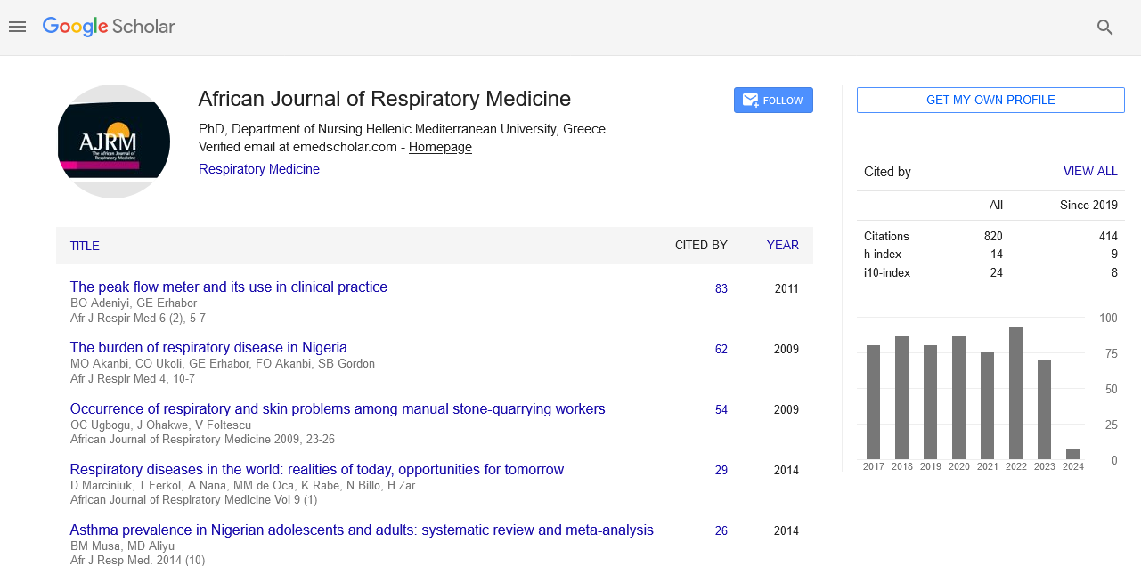
African Journal of Respiratory Medicine received 855 citations as per google scholar report
Case Report - (2024) Volume 19, Issue 2
Received: 07-Mar-2024, Manuscript No. ajrm-24-129107; Editor assigned: 11-Mar-2024, Pre QC No. ajrm-24-129107(PQ); Reviewed: 25-Mar-2024, QC No. ajrm-24-129107; Revised: 01-Apr-2024, Manuscript No. ajrm-24-129107 (R); Published: 08-Apr-2024, DOI: 10.54931/1747-5597.24.19.02
South Africa has a high burden of tuberculosis (TB) and it is the leading cause of death in the country. Pulmonary embolism (PE) and Pulmonary TB can share many of the same symptoms and can coexist in the same patient, making the diagnosis challenging. Furthermore, venous thromboembolism (VTE) is more common among patients with TB compared to the general population. Here we describe three cases of patients with Pulmonary TB, on treatment, complicated by PE. Our cases highlight the need for a high index of suspicion for PE in patients with severe TB.
Tuberculosis; Pulmonary embolism; Venous thromboembolism
Tuberculosis (TB) and pulmonary embolism (PE) are two conditions that affect the respiratory system and can share similar symptoms.1 Both can coexist in the same patient, making diagnosis challenging.1 TB is a major public health concern in South Africa (SA) and the nation continues to have a high incidence and prevalence of TB.2,3 The prevalence, in a 2017 national survey, was 737/100,000 with an incidence of 567 cases per 100,000 populations.3,4 While effective treatment for TB is widely available it remains the principal cause of death in SA.3,5 Venous Thromboembolism (VTE) is categorised as PE and Deep Venous Thrombosis (DVT) and is correlated with large financial burdens on the health system and high morbidity on patients.6 PE is a life threatening condition and is associated with in-patient mortality as high as 23%.7 A meta-analysis from 2020 found that the prevalence of VTE in patients with active TB was 4 to 8 times higher compared to the general population.5 Moreover, PE was found to have a prevalence of 5.8% in these patients.5 Here we describe PE in three patients with severe TB at Rob Ferreira Hospital and their outcomes.
Case 1
A 43-year-old male diagnosed with pulmonary TB, already on intensive phase of Anti-tuberculous Treatment (ATT), presented to our hospital with a three day history of shortness of breath, a non-productive cough, and retrosternal chest pain. Physical examination revealed the following: Blood pressure 112/87 mmHg, pulse 130 bpm, respiratory rate 40 bpm, pulse-oximetry 80% on room air and 100% on high flow nasal cannula oxygen therapy. No significant lymphadenopathy was present. A diffuse reticulonodular pattern was evident on chest roentgenogram. After one week of in-patient treatment with no improvement and taking into account all findings, there was a high index of suspicion for pulmonary embolism. The only risk factor for PE being that the patient was non-ambulatory due to his illness. A Computed Tomography Pulmonary Angiogram (CTPA) confirmed PE in branches of right pulmonary artery (Figure 1). The patient was initiated on therapeutic doses of enoxaparin and anticoagulated with warfarin. The patient was discharged after a 15-day hospital stay.

Figure 1. Doppler ultrasound demonstrating right common femoral DVT (white asterisk)
Case 2
A 36-year-old male with Human Immunodeficiency Virus (HIV) diagnosed with pulmonary TB, on intensive phase of ATT, presented to our hospital with a three week history of progressive swelling and pain of his right lower limb. Physical examination showed the following: Blood pressure 146/70 mmHg, pulse 111 bpm, respiratory rate 20 bpm, pulse-oximetry 98% on room air. No lymphadenopathy was palpable on examination. Local examination of the lower limbs showed a unilateral swollen right lower limb with tenderness to deep palpation. Ultrasound Doppler of the lower limbs (Figure 2) revealed a right ileofemoral DVT. The patient was initiated on therapeutic enoxaparin and anticoagulated with warfarin. However, during the second day of the patient’s hospital admission he developed severe type 1 respiratory failure and pleurisy. A CTPA revealed bilateral PE (Figure 3). The only identifiable risk factor for PE was current DVT. A decision was made to give the patient thrombolysis due to the extensive nature of the thromboembolic disease. The patient’s hypoxia resolved following thrombolysis and the patient was discharged after a 7-day hospital stay.

Figure 2. Doppler ultrasound demonstrating right common femoral DVT (white asterisk)

Figure 3. Axial CTPA demonstrating PE in right pulmonary artery and its branches and in left pulmonary artery branches (white arrows)
Case 3
A 50-year-old male with HIV diagnosed with disseminated TB, on continuation phase of ATT, presented with progressive weakness of his left upper limb and both lower limbs and pleurisy for the last month. Physical examination showed the following: Blood pressure 97/69 mmHg, pulse 114 bpm, respiratory rate 16 bpm, and pulse-oximetry of 96%. Examination revealed a chronically ill-patient with generalised lymphadenopathy, an asymmetrical sensorimotor neuropathy and clinical features of pulmonary hypertension. Point of care ultrasound showed moderate tricuspid regurgitation with a pulmonary artery systolic pressure of 51 mmHg, dilated right heart chambers, and dilated non-collapsing inferior vena cava. On review of all findings there was a high index of suspicion for PE. The only apparent risk factor for PE was that the patient was bedridden due to his neuropathy. CTPA revealed bilateral PE (Figure 4). The patient was initiated on therapeutic doses of enoxaparin and anticoagulated with warfarin. Patient was discharged form hospital after a 38-day hospital stay.

Figure 4. Axial CTPA demonstrating pulmonary embolism in right pulmonary artery and its branches (white arrow)
Even though TB is the main cause of death in SA, PE is infrequently described as a complication of TB.8 TB increases the probability of VTE by increasing the likelihood of hypercoagulability, stasis, and endothelial lesions.5 Patients with severe TB are often bedridden for extended periods of time, which raises their risk for PE.5 Furthermore, TB with pulmonary lymph node enlargement can cause direct venous compression leading to thrombosis.1,5 TB can cause endothelial damage with resultant release of pro-coagulants such as kallikrein and activation of the complement cascade further promoting VTE.1 Additionally, the risk of VTE in TB is amplified by reduced anti-thrombin III, protein C and increased factor VIII, plasminogen activator inhibitor I, and fibrinogen serum levels.1 There is a substantial increase of anti-phospholipid antibodies that have been shown to have a dose dependent elevation in the risk of VTE.9 Moreover, the hypercoagulable state is worsened by thrombocytosis and disrupted fibrinolysis in severe TB.1 Treatment of TB with rifampicin and isoniazid contributes to this response by increasing anticoagulant clearance and disruption of coagulation factors.1 Rifampicin has also been linked to VTE by immunological processes.10 TB is major health concern in South Africa and there is evidence to suggest that TB increases the risk of developing PE.3,5 Our cases emphasise the significance of a high index of suspicion for PE in patients with severe TB.
TB is major health concern in South Africa and there is evidence to suggest that TB increases the risk of developing PE. Furthermore TB is the leading cause of natural death in SA. It is important for primary health care physicians to be aware of this life threatening complication of a common and dangerous disease entity. Our cases emphasise the significance of a high index of suspicion for PE in patients with severe TB.
None.
None declared.
None declared.
Arthur (AE) McKinnon is the guarantor for this manuscript.
[Crossref] [PubMed] [Google Scholar]
[Crossref] [PubMed] [Google Scholar]
[Crossref] [PubMed] [Google Scholar]
[Crossref] [PubMed] [Google Scholar]
[Crossref] [PubMed] [Google Scholar]
[Crossref] [PubMed] [Google Scholar]
[Crossref] [PubMed] [Google Scholar]
Select your language of interest to view the total content in your interested language
To read the issue click on a cover